Retina
What is the retina?
The retina is the back layer of the eye and is made up of cells that are sensitive to light. The cells turn light into nerve impulses, which travel through the optic nerve to the brain, where they are interpreted as images. The most sensitive part of the retina is an area known as the macula, which is responsible for high-resolution images (mainly cone cells).

Regular check-ups can prevent the development of diseases related to the retina, or can stop them getting worse.
Retinal disorders
Retinopathy secondary to other conditions such as diabetes or arterial hypertension.
Age-related Macular Degeneration (AMD), the most common cause of progressive loss of vision in the elderly. In advanced cases, it will appear as a stain in the centre of the patient’s vision accompanied by distorted images. There are two types of AMD:
- Dry or atrophic AMD, which involves progressive cell atrophy and/or a build-up of waste materials (drusen).
- Wet or exudative AMD, which is related to the formation of new blood vessels (neovascular membranes). This can cause a build-up of fluid or haemorrhages between the retinal layers.
Vascular involvement due to thrombosis or embolism, which can affect either a branch of the veins in the eye, or all of them.
Central serous chorioretinopathy, which appears abruptly and affects central vision. This conditions is associated with people who suffer from nerves and stress.
Infections: viral infections (herpes virus) or parasitic infections (toxoplasmosis, toxocariasis).
Retinal detachments with different causes, although they are most commonly started by a tear or rupture of the retina.
Multiple congenital degenerative disorders, such as retinitis pigmentosa or Stargardt disease. Many of these conditions are hereditary.

Normal retina.

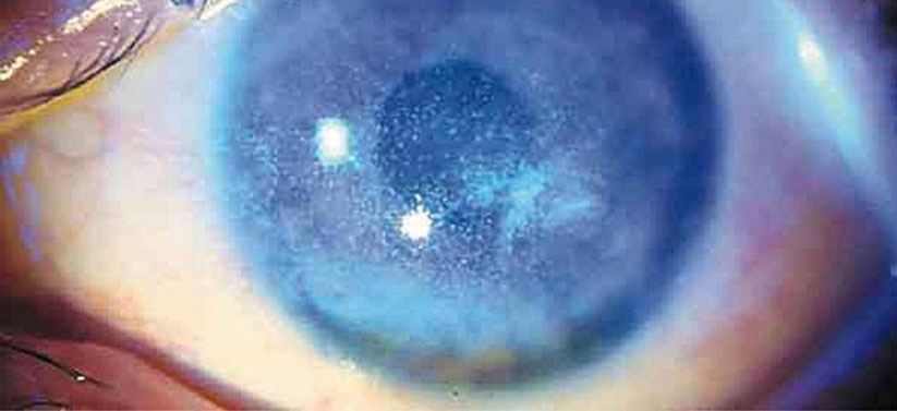
Dry AMD: Areas of chorioretinal atrophy and alteration of the retinal pigment epithelium in the macula.


Central field of vision is affected by AMD.
Evaluation
Diagnostic techniques
Visual acuity (with best-corrected visual acuity).
Campimetry: This test allows us to detect alterations in both the central and peripheral field of vision.
Dilated fundus exam, dilating the pupil for a full examination of the retina.
Technological instruments
Retinography: Photograph of the retina.
Optical coherence tomography (OCT): This state-of-the-art instrument detects microscopic retinal alterations even before the patient starts to show symptoms.
External tests:
- Fluorescein angiography and indocyanine green angiography: This test examines the condition of the eye’s blood vessels using a contrast dye.
- Electrophysiological tests: Electroretinogram tests, visual evoked potentials and electro-oculogram tests, among others.

Normal state of the macula, as seen with OCT.

On the left, wet AMD with oedema and macular haemorrhage. On the right, OCT showing a build-up of fluid under the layers of the retina (see arrow) and showing the neovascular membrane (see asterisk).
A serous detachment of the neurosensory retina is shown as a result of a central serous chorioretinopathy.
OTHER SPECIALITIES
Treatment of retinal diseases
- In the case of dry AMD, introducing antioxidants in the patient’s diet can slow down its progression. For wet AMD, there are certain drugs that can be injected into the eye, removing the fluid build-up in the retina and preventing it from coming back, allowing vision to stabilise and improve.
- Diseases related to metabolic imbalances can be controlled by a good diet and medication (as for diabetes or hypertension). Monitoring cholesterol levels and exercising can also prevent thrombosis or embolism.
- Central serous chorioretinopathy has a good prognosis and usually resolves itself simply by avoiding the stress that caused it.
- Some infections are caused by coming into contact with certain animals (toxoplasmosis), standing water or raw meat.
- Retinal tears can be caused by blows to at-risk patients, such as those with high degree myopia. If caught in time, they can be treated with laser photocoagulation.
- Unfortunately, some retinal diseases cannot be cured (such as retinitis pigmentosa), although future treatments are being investigated.

Adding antioxidants to your diet can help slow the progression of the dry type of age-related macular degeneration.
Frequently Asked Questions
Your eyes are full of a gel called vitreous. This can form different kinds of “deposits” that will sometimes project their shadow on the retina, which is then seen as “floaters”. With age, the vitreous can detach and many can appear at once. If this happens, you should book an appointment to rule out the possibility of retinal tears.
- Seeing “flashes of light” together with “floaters” anywhere in your field of vision.
- An unmoving dark patch anywhere on your field of vision that does not disappear when blinking.
- Seeing an unmoving spot or “wavy lines” in the central part of your field of vision. Sudden and generally painless loss of vision.
Related entries
Solar radiation and eye health
Solar radiation affects our eye health more than we can imagine. Solar rays are formed not only by the visible…